![]() Figure 4 of
Jin, Mol Vis 2007;
13:626-634.
Figure 4 of
Jin, Mol Vis 2007;
13:626-634.
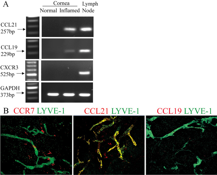
Figure 4. CCR7 and CCL21 were expressed around neo-lymphatic vessels in the vascularized corneas
Transcript of CCL21, but not CCL19 or CXCR3, was significantly expressed in the inflamed corneas. Lymph nodes served as positive control (A). In the inflamed corneal stromas, dendritiform CCR7+ cells (left, red) and CCL21+ cells (middle, red) were near the LYVE-1+ lymphatics (green). CCL21 also co-localized with LYVE-1+ lymphatics (middle, yellow). No CCL19+ cells were detected (right; B). Confocal microscopy, original magnification: X40.