![]() Figure 3 of
Jin, Mol Vis 2007;
13:626-634.
Figure 3 of
Jin, Mol Vis 2007;
13:626-634.
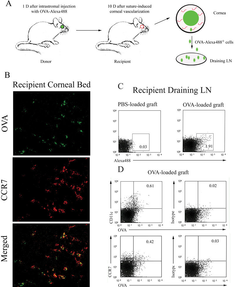
Figure 3. CCR7 was expressed on OVA+ cells in both the recipient corneal beds and the draining lymph nodes
OVA-loaded corneal grafts were transplanted into vascularized syngeneic recipient beds (A). After 48 h, a portion of CCR7+ cells (red) also expressed OVA (green) on their surface in the recipient stromal beds (costained as yellow). Confocal microscopy, original magnification: X40 (B). Draining lymph node (LN) cells were harvested and OVA+ cells were detected by flow cytometry. Mice with PBS-loaded corneal grafts served as negative controls (C). Flow cytometric expression of CD11c and CCR7 on the OVA+ cells in the draining LN is shown in D.