![]() Figure 4 of
Wang, Mol Vis 2007;
13:618-625.
Figure 4 of
Wang, Mol Vis 2007;
13:618-625.
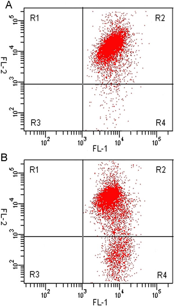
Figure 4. Analysis of Δψm in human trabecular meshwork cells
A: The scattergram showed the most cells presented in the R2 region with high red fluorescence (FL-2) and high green fluorescence (FL-1), which revealed normal Δψm of human trabecular meshwork (HTM) cells. B: HTM cells transfected with Pro370Leu mutant myocilin plasmid. The cells markedly decreased in the R2 region, indicating that Pro370Leu mutant myocilin induced loss of Δψm. A representative experiment of three is shown.