![]() Figure 3 of
Suganthalakshmi, Mol Vis 2007;
13:611-617.
Figure 3 of
Suganthalakshmi, Mol Vis 2007;
13:611-617.
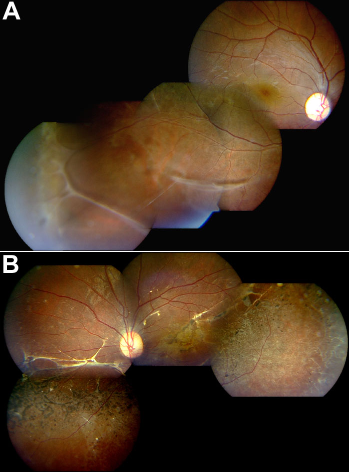
Figure 3. Fundus photography of patient 5 with X-linked juvenile retinoschisis
A: Composite fundus photographs of the right eye of patient 5 showing foveal schisis, as well as peripheral vitreous veils. B: Composite fundus photograph of the left eye of the same patient, showing spontaneously settled inferotemporal retinal detachment involving macula, with consequent foveal atrophy.