![]() Figure 2 of
Suganthalakshmi, Mol Vis 2007;
13:611-617.
Figure 2 of
Suganthalakshmi, Mol Vis 2007;
13:611-617.
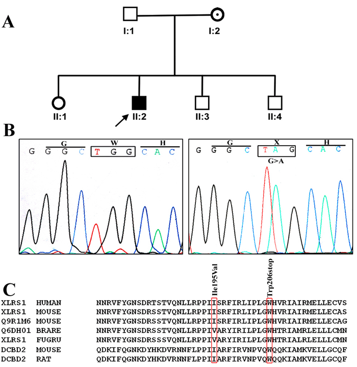
Figure 2. Genetic analysis of patient 4 with X-linked juvenile retinoschisis
A: Family pedigree of patient 4, who exhibited a novel c.617G>A mutation. The arrow indicates proband. The shaded box denotes that the proband affected with retinoschisis. The dotted circle indicates carrier. B: This sequence chromatogram compares the novel c.617G>A nonsense mutation with the sequence derived from control. The DNA of proband II: 2 revealed a homozygous G to A transition in exon 6 of RS1 gene that leads to the replacement of aminoacid tryptophan to stop codon at position 206. C: A multiple sequence alignment for I195V and W206X novel genetic variants of RS1 gene, in different species and compared with other retinoschisin and discoidin domain proteins. The amino acids boxed in red indicate the position of I195V, and W206X. The aminoacid tryptophan is highly conserved among the various species.
