![]() Figure 1 of
Suganthalakshmi, Mol Vis 2007;
13:611-617.
Figure 1 of
Suganthalakshmi, Mol Vis 2007;
13:611-617.
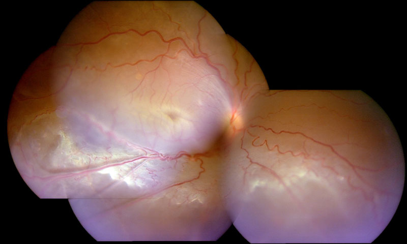
Figure 1. Fundus photography of patient 4 with X-linked juvenile retinoschisis
Composite fundus photograph of the right eye of patient 4 showing total rhegmatogenous retinal detachment secondary to inferotemporal schisis, which is evident as elevated inferotemporal retinal vessels (vitreous veils). The foveal schisis is obscured by the detachment.