![]() Figure 3 of
Shimazawa, Mol Vis 2007;
13:578-587.
Figure 3 of
Shimazawa, Mol Vis 2007;
13:578-587.
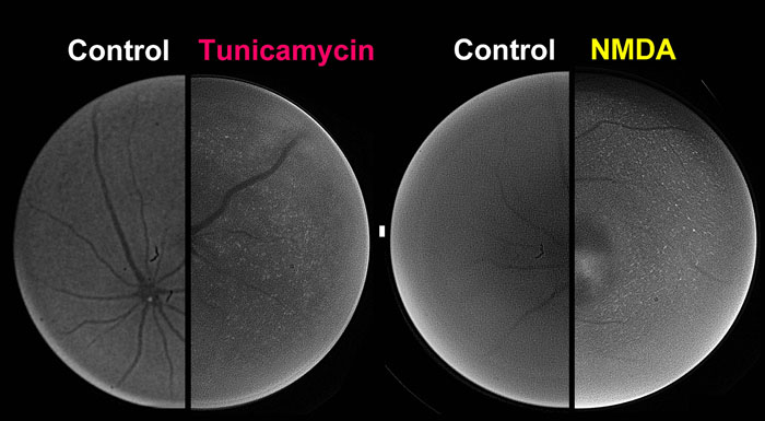
Figure 3. Non-invasive imaging of XBP-1-venus fusion protein in ERAI mouse retina in vivo
Twenty-four hours after intravitreal injection of either tunicamycin at 0.1 μg/eye or N-methyl-D-aspartate (NMDA) at 40 nmol/eye, the fluorescence intensity arising from XBP-1-venus fusion protein was visualized in the retinas of anesthetized animals using an ophthalmoscope fitted with a fluorescence filter.