![]() Figure 6 of
Yaung, Mol Vis 2007;
13:566-577.
Figure 6 of
Yaung, Mol Vis 2007;
13:566-577.
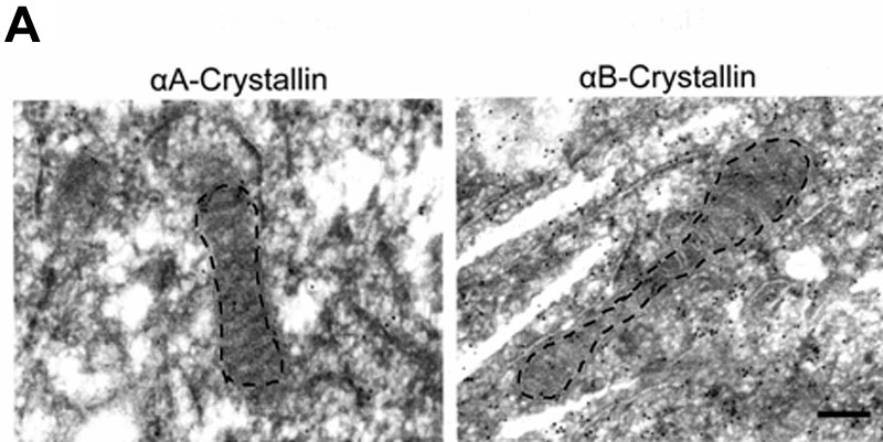
Figure 6. Immuno-transmission electron microscopy of gold-conjugated α-crystallin in human retinal pigment epithelium
Retinal pigment epithelium (RPE) cells treated with and without H2O2 were pelleted, fixed, sectioned and stained with 10 nm gold particles conjugated with α-crystallin antibody. Representative micrographs of untreated mitochondria in RPE labeled for αA (left) and αB-crystallin (right) are shown (A). Quantitation of gold particles in mitochondria revealed a dose-dependent decrease of crystallins; however, only αB-crystallin demonstrated statistical significance compared to untreated controls (B). Due to the variability of mitochondria size in sectioned samples, gold particle counts were normalized to untreated controls. An average of 15 micrographs was quantited for each condition. Asterisks indicate p<0.05 versus untreated controls. The scale bar represents 0.5 μm.