![]() Figure 2 of
Yaung, Mol Vis 2007;
13:566-577.
Figure 2 of
Yaung, Mol Vis 2007;
13:566-577.
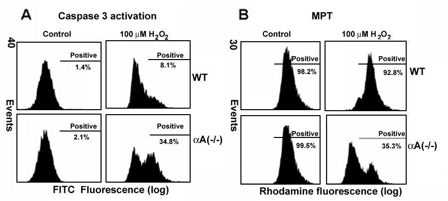
Figure 2. Effect of H2O2 treatment in retinal pigment epithelium from αA-crystallin(-/-) and wild type mice on caspase-3 activation and mitochondrial membrane permeability transition
A: Caspase-3 activation was measured by staining with FITC-VAD-FMK. αA(-/-) retinal pigment epithelium (RPE) showed an increase in caspase-3 activation compared to wild type under H2O2 treatment. B: αA(-/-) RPE also exhibited an increase in mitochondrial permeability transition compared to wild type RPE under H2O2 treatment.