![]() Figure 1 of
Yaung, Mol Vis 2007;
13:566-577.
Figure 1 of
Yaung, Mol Vis 2007;
13:566-577.
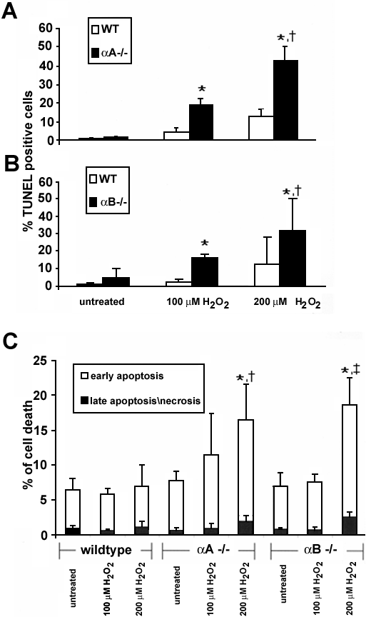
Figure 1. Effect of H2O2 on cell death in retinal pigment epithelium from α-crystallin (-/-) and wild type mice
RPE from knockout mice grown to confluency were treated with 100 μM or 200 μM H2O2 for 24 h and stained for apoptosis by TUNEL and quantified by flow cytometry. Cell death of RPE increased in a dose dependent manner; however, αA(-/-) (A) and αB(-/-) (B) RPE showed an increased sensitivity to apoptosis from H2O2 treatment compared to wild type cells. In the α-crystallin knockout RPE at the maximum oxidative stress used (200 μM H2O2), >85% of the dead cell population was AnnexinV+/PI- indicating an apoptotic mechanism of cell death; <15% of cells were positive for both Annexin V and PI indicating either necrosis or later stage apoptosis (C). Data are mean±SD (n=3-4). One and two asterisks indicate p<0.05 for the two H2O2 doses versus untreated wild type controls, and p<0.05 for dose-dependency in TUNEL positive cells in knockout mice at 100 μM and 200 μM H2O2 doses, respectively. Difference between apoptosis in untreated αA(-/-) and αB(-/-) RPE compared to wild type did not achieve statistical significance (p=0.25).