![]() Figure 9 of
Montiani-Ferreira, Mol Vis 2007;
13:553-565.
Figure 9 of
Montiani-Ferreira, Mol Vis 2007;
13:553-565.
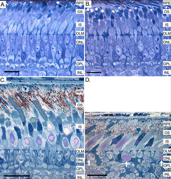
Figure 9. Semithin sections of the outer retina of control and rge birds at one and 270 days of age
Semithin retinal sections showing the outer retina of a control (A) and rge (B) chick at one day of age and a control (C) and rge (D) chicken at 270 days of age. The 1-day-old rge retina had only subtle morphological retinal abnormalities, involving slight dilation of inner segments and some disorganization of the outer plexiform layer. The affected birds had a slow thinning of the photoreceptor layer with age. By 270 days of age the rge bird had a marked thinning of the photoreceptor layer and further disruption of the outer plexiform layer. There was also the presence of displaced glycogen deposits adjacent to photoreceptor nuclei (arrows). Photomicrographs adapted with permission from: Montiani et al 2005 [15]. Size bars for A and B represent 10 μm. Size bars for C and D represent 20 μm. RPE indicates retinal pigment epithelium; OS indicates photoreceptor outer segments; IS indicates photoreceptor inner segments; OLM indicates outer limiting membrane; ONL indicates outer nuclear layer; OPL indicates outer plexiform layer; INL indicates inner nuclear layer.