![]() Figure 3 of
Rutland, Mol Vis 2007;
13:47-56.
Figure 3 of
Rutland, Mol Vis 2007;
13:47-56.
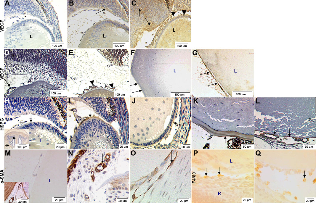
Figure 3. Immunohistochemical detection of VEGF, HSPG, α-SMA, and F4/80 in eyes from wild-type and VEGF-A transgenic mice
Immunohistochemical detection of VEGF-A in wild-type and transgenic mouse eyes at E15.5 (A-C), P1 (D+E), and adult (F-G). A; Non-specific IgG serum (control), no discernible background staining is observed. B: VEGF-A immunostaining is observed in the wild-type lens (L) and superficial layers of the retina (black arrow) at E15.5. However specific VEGF-A staining was not detected at P1 in the retina, lens, or hyaloid vasculature (D). In the adult wild-type eye (F), specific staining of the surface of the lens capsule is seen (arrows). Specific staining for VEGF-A is observed in the lens (L), retina (black arrows) and adjacent to the hyaloid vasculature (black arrowheads) at both E15.5 (C) and at P1 (E) in VEGF-A188 transgenics. In adult VEGF-A188 mice (G), prominent staining is observed in the lens capsule (arrows) which appears thinner and (during processing) has separated from the lens structure. Immunohistochemical detection of HSPG (H-L), α-SMA (M-O), and F4/80 (P,Q) in sections from E15.5 (H-J, M-Q) and adult (K,L) mice are shown. H: No discernible background staining is observed in control sections (IgG-specific serum). Inset shows an E15.5 mouse brain section, which served as a positive control. I: In wild-type mice, specific HSPG staining is observed in the lens capsule and hyaloid vasculature (arrow). J: In VEGF-A188 transgenic mice, HSPG staining is observed in the lens capsule (arrow). K: HSPG staining is also observed in lens capsule of adult wild-type mice (arrow), with a characteristic laminar pattern. L: In adult VEGF-A188 transgenic mice, HSPG staining is seen in the thin lens capsule (arrow) and around persistent hyaloid vasculature (open arrows). Conspicuous staining of peri-vascular smooth muscle cells surrounding blood vessels (arrow) in both E15.5 wild-type (N) and transgenic eyes (O) was noted. F4/80 immunoreactivity in sections from E15.5 wild- type (P) and transgenic mouse eyes (Q) revealed cells adjacent to the hyaloid vasculature. In the images, L=lens, R=retina, HV=hyaloid vasculature.