![]() Figure 1 of
Rutland, Mol Vis 2007;
13:47-56.
Figure 1 of
Rutland, Mol Vis 2007;
13:47-56.
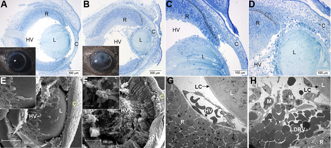
Figure 1. Gross anatomical and microscopic features of VEGF-A188 transgenic mice
Representative photomicrographs of toluidine blue-stained sections from E18.5 wild-type (A,C) and VEGF-A188 transgenic (B,D) mice. Gross ocular phenotypes of adult wild-type (A; inset) and a VEGF-A188 transgenic mouse (B; inset), showing conspicuous cataract formation. In VEGF-A188 transgenic mice (B, D), a hypertrophic hyaloid vasculature surrounds a small lens. There is evidence of retinal hypertrophy, particularly in the ganglion cell layer at the top of these micrographs. Scanning electron micrographs of wild-type (E) and VEGF-A188 transgenic (F) mice are shown. The thickened hypertrophic hyaloid vasculature with numerous adherent mononuclear cells in the VEGF-A188 transgenic lens (F) contrasts with the organized plexus in wild-type mice. Transmission electron micrographs of hyaloid blood vessels in E18.5, wild-type (G), and VEGF-A188 transgenic mice (H). Large diameter vessels(LDBV), with several attendant macrophages (M), are conspicuous in VEGF-A188 transgenics. In the images, C=cornea, HV=hyaloid vasculature, R=retina, and L=lens.