![]() Figure 6 of
Proulx, Mol Vis 2007;
13:524-533.
Figure 6 of
Proulx, Mol Vis 2007;
13:524-533.
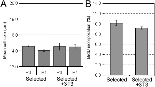
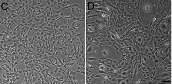
Figure 6. Effect of coculturing porcine corneal endothelial cells (PCEC) with a feeder layer of irradiated 3T3 cells grown in our selected medium
A: Mean cell size of PCEC cultured at different passages (P) after seeding at the very low cell density of 400 cells/cm2 without a feeder layer (Selected) and with a feeder layer (Selected+3T3). B: Percentage of bromodeoxyuridine (BrdU) incorporation of PCEC seeded at 400 cells/cm2 at P1 cultured without a feeder layer (Selected) and with a feeder layer (Selected+3T3; mean±SD). The student's t-test performed shows that there is no statistically significant difference between P0 and P1 (p<0.001). Morphology of PCEC (P1) seeded at 400 cells/cm2 and cultured without a feeder layer (C) and with a feeder layer (D), both grown in the selected medium, consisting of Opti-MEM I supplemented with 8% fetal bovine serum (FBS), 50 μg/ml bovine pituitary extract (BPE), 0.08% chondroitin sulfate and 20 μg/ml ascorbic acid. The scale bar is equal to 100 μm.