![]() Figure 5 of
Proulx, Mol Vis 2007;
13:524-533.
Figure 5 of
Proulx, Mol Vis 2007;
13:524-533.
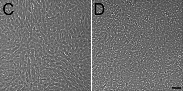
Figure 5. Effect of different culture media formulations on cell number, size, and morphology of porcine corneal endothelial cells (PCEC)
Representative results of the additive effect of 50 μg/ml bovine pituitary extract (BPE), 0.08% chondroitin sulfate (0.08 CDS) and 20 μg/ml ascorbic acid (20AA) on (A) mean cell size and (B) cell number at day 4 on PCEC grown in Opti-MEM I supplemented with 0, 4, or 8% fetal bovine serum (FBS), and compared to the classic Dulbecco's Modified Eagle's Medium (DMEM) 20% FBS (dashed bar graph; mean±SD). The asterisk indicates a p<0.001 compared to DMEM 20% FBS (cell number and cell size). Morphology of PCEC grown in C DMEM 20% FBS or D the selected medium, consisting of Opti-MEM I supplemented with 8% FBS, 50 μg/ml BPE, 0.08% chondroitin sulfate and 20 μg/ml ascorbic acid. Cells were left in culture one week passed confluence to assess cell morphology of post-confluent cultures. The scale bar is equal to 100 μm. Note that in (C) endothelial cells are elongated and of different size whereas in (D) they are small and cuboidal cells and have a morphology more characteristic of native cells.