![]() Figure 4 of
Proulx, Mol Vis 2007;
13:524-533.
Figure 4 of
Proulx, Mol Vis 2007;
13:524-533.
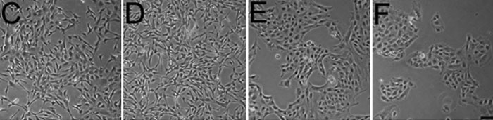
Figure 4. Effect of different additives on cell number, size, and morphology of porcine corneal endothelial cells (PCEC)
Representative results of the dose-dependent effect of various growth-promoting factors. PCEC were cultured in Opti-MEM I 4% fetal bovine serum (FBS) and one of the following factors: epidermal growth factor (EGF; 0.5, 5, and 25 ng/ml), nerve growth factor (NGF; 5, 20, and 50 ng/ml), bovine pituitary extract (BPE; 25, 50, 100, and 200 μg/ml), chondroitin sulfate (CDS; 0.03, 0.08, and 1.6%) and ascorbic acid (AA; 10, 20, 40 μg/ml). A shows the mean cell size. B: Number of cells/cm2. The results are the mean±SD. The asterisk indicates a p<0.001 compared to Opti-MEM I 4% FBS (cell number and cell size). Morphology at day 4 of PCEC grown in C BPE 50 μg/ml, D BPE 200 μg/ml, E ascorbic acid 20 μg/ml, and F chondroitin sulfate 0.08%. The scale bar is equal to 100 μm.