![]() Figure 2 of
Proulx, Mol Vis 2007;
13:524-533.
Figure 2 of
Proulx, Mol Vis 2007;
13:524-533.
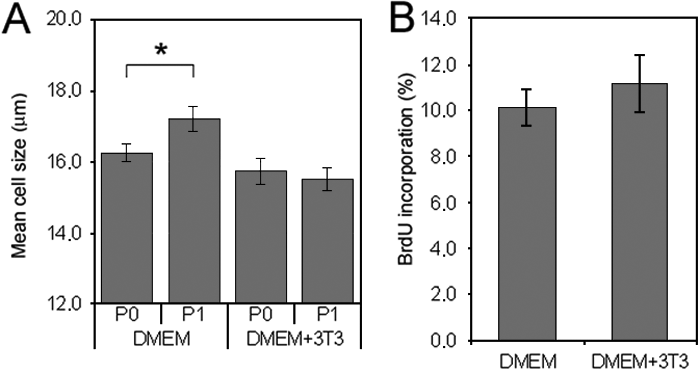
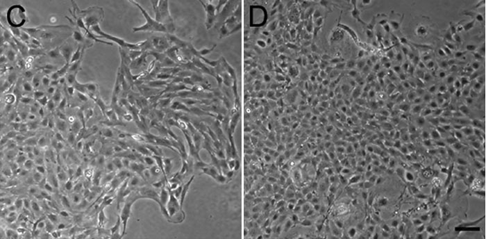
Figure 2. Effect of coculturing porcine corneal endothelial cells (PCEC) with a feeder layer of irradiated 3T3 cells grown in Dulbecco's Modified Eagle's Medium (DMEM) 20% fetal bovine serum (FBS)
A: Mean cell size of PCEC cultured at different passages (P) after seeding at the very low cell density of 400 cells/cm2 without a feeder layer (DMEM) and with a feeder layer (DMEM+3T3). B: Percentage of bromodeoxyuridine (BrdU) incorporation of PCEC seeded at 400 cells/cm2 at P1 cultured without a feeder layer (DMEM) and with a feeder layer (DMEM+3T3). The results are the mean±SD. Student t-test performed between P0 and P1 (the asterisk indicates a p<0.001). Morphology of PCEC (P1) seeded at 400 cells/cm2 and cultured without a feeder layer (C) and with a feeder layer (D). The scale bar equals 100 μm. Note the smaller size of porcine endothelial cells in the presence of a feeder layer.