![]() Figure 1 of
Proulx, Mol Vis 2007;
13:524-533.
Figure 1 of
Proulx, Mol Vis 2007;
13:524-533.
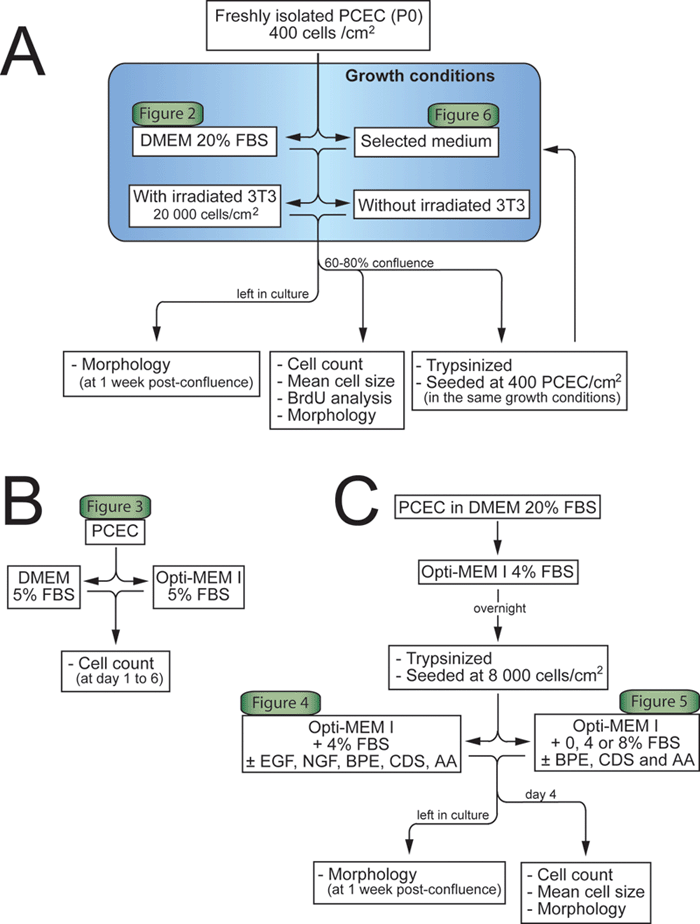
Figure 1. Flow diagram of the experimental protocol.
A: Effect of coculture. Freshly isolated porcine corneal endothelial cells (PCEC) in primary culture (P0) seeded at 400 cells/cm2 were cultured in Dulbecco's Modified Eagle's Medium (DMEM) 20% fetal bovine serum (FBS) or in the selected medium with or without a feeder layer of irradiated murine 3T3 cells. Cells were trypsinized at 60-80% confluence and subcultured in the same conditions. Cell number, mean cell size, and bromodeoxyuridine (BrdU) analysis were assessed on the pre-confluent cells. Other cultures were maintained one week after confluence to document their morphology at post-confluence. B: Effect of basal media. PCEC were seeded at 12,000 cells/cm2 either in DMEM or Opti-MEM I, both supplemented with 5% FBS. Cells of each well were counted from day 1 to day 6. C: Effect of growth promoting additives. PCEC seeded at 8,000 cells/cm2 were cultured in DMEM 20% FBS and switched to Opti-MEM I 4% FBS overnight. PCEC were then trypsinized and plated at 8,000 cells/cm2 and cultured in the presence of the following additives (used alone or in combination): epidermal growth factor (EGF), nerve growth factor (NGF), bovine pituitary extract (BPE), ascorbic acid (AA), and chondroitin sulfate (CDS). Cell number, size, and morphology were assessed on day 4. Also, the selected medium containing BPE, CDS, and AA was tested in the presence (4% and 8%) or absence of serum. See the methods section for further details.
