![]() Figure 6 of
Dansault, Mol Vis 2007;
13:511-523.
Figure 6 of
Dansault, Mol Vis 2007;
13:511-523.
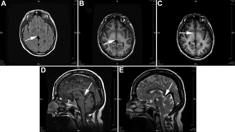
Figure 6. Brain MRI abnormalities in patient IV-5 from family F with the PAX6 serine 74 to glycine mutation
We show different MRI views of patient IV-5 from family F. A, B, and C correspond to MRI horizontal views, while D and E correspond to MRI sagittal views. MRI in A shows a small structure that might correspond to the habenula (arrow). MRI in B, D, and E reveals the absence of the pineal gland (arrow). MRI in C shows hypoplasia of the anterior commissure of the brain (arrow).