![]() Figure 4 of
Dansault, Mol Vis 2007;
13:511-523.
Figure 4 of
Dansault, Mol Vis 2007;
13:511-523.
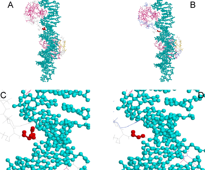
Figure 4. Crystallographic views of the serine 74 to glycine mutation found in family F
Represented are the interactions between the DNA (blue) and PAX6 PD red. A and B show global views while C and D represent magnified views of the linker region of the PD (residue 61-76) in which the mutated Ser74 is located. This region makes many contacts with the minor groove of the DNA strand. In the wild-type protein (A and C), the Ser-74 (red) side chain contacts the DNA. In the mutated protein (B and D), the amino acid change leads to the loss of the side chain and increases the distance between the target genomic DNA and the PAX6 linker domain, making direct contact between DNA and PAX6 impossible.