![]() Figure 3 of
Dansault, Mol Vis 2007;
13:511-523.
Figure 3 of
Dansault, Mol Vis 2007;
13:511-523.
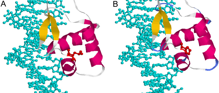
Figure 3. Crystallographic views of leucine 46 to proline mutation found in family E
Represented are the interactions between DNA (blue) and the helix-turn-helix motif affected by the amino acid change (red) in the wild-type protein (A) and in the mutated protein (B). The mutated Leu 46 is located just after the second helix in the NH2-terminal subdomain of the PAX6 paired domain (PD). The substitution of a proline for a leucine modifies the structure of the second helix and affects the HTH motif conformation. This could alter the position of the third helix, which recognizes the DNA. The readers should be aware that we standardized the numbering of the amino acid residues of the paired domain. We allocated a number to each amino acid residue that corresponded to its location vis-a-vis the first methionine, initiating the PAX6 short protein isoform (-5a). However, the X-ray crystallography specialists use to allocate a numbering to the amino acids of the PAX6 PD which starts at the first amino acid of this PD.