![]() Table 1 of
Meng, Mol Vis 2007;
13:475-486.
Table 1 of
Meng, Mol Vis 2007;
13:475-486.
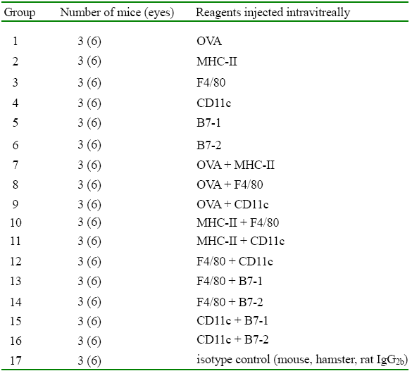
Table 1. Groups designed for in vivo immunofluorescent staining
To visualize the labeled cells in the cornea, fifty-one BALB/c mice were divided into 17 groups and 2 μl of fluorescently tagged OVA or antibodies were injected into these murine vitreous bodies, respectively, according to designed combinations. Except for OVA, all were antibodies against the indicated cell surface molecules.
