![]() Figure 9 of
Meng, Mol Vis 2007;
13:475-486.
Figure 9 of
Meng, Mol Vis 2007;
13:475-486.
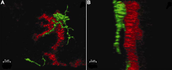
Figure 9. Three-dimensional rotatable confocal microscopy images for observing the relationship between labeled cells
The MHC-II+ cell contacts an OVA+ cell at a portion of the cell body and the dendrite (A: the frontal image, B: the lateral image; red OVA, green MHC-II).