![]() Figure 8 of
Meng, Mol Vis 2007;
13:475-486.
Figure 8 of
Meng, Mol Vis 2007;
13:475-486.
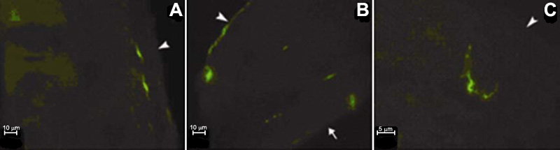
Figure 8. Location of labeled cells in the vertical axis of the cornea
Frozen sections are prepared and stained with antibodies and OVA, followed by epifluorescence microscopy examination. OVA+ cells are distributed beneath the epithelium in the central part (A) and present in all layers of the peripheral cornea (B). The dendrites projecting from MHC-II+ cells are present longitudinally into the epithelium and stroma (C). Arrowheads indicate the epithelium location and the arrow indicates the location of endothelium.