![]() Figure 7 of
Meng, Mol Vis 2007;
13:475-486.
Figure 7 of
Meng, Mol Vis 2007;
13:475-486.
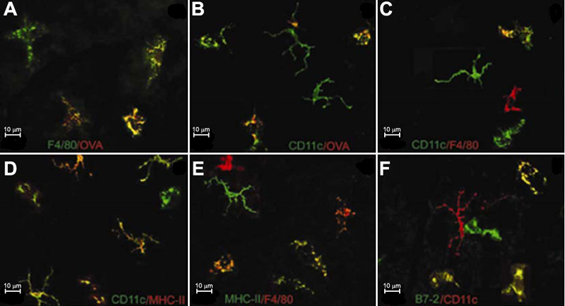
Figure 7. Phenotypes of APCs in the normal cornea shown in confocal microscopy images
Corneas are double stained in vivo with OVA and antibodies, followed by examination of wholemounts under the confocal microscopy. A number of F4/80+ (A: green) and CD11c+ (B: green) cells are positive for OVA (A,B: red). A few of F4/80+ cells are positively costained with anti-CD11c (C: red F4/80, green CD11c). A number of CD11c+ cells (D: green) and F4/80+ cells (E: red) are also MHC-II positive (D: red, E: green). More than half of CD11c+ cells are double stained for B7-2 (F: red CD11c, green B7-2). Double-labeled cells are yellow.