![]() Figure 6 of
Meng, Mol Vis 2007;
13:475-486.
Figure 6 of
Meng, Mol Vis 2007;
13:475-486.
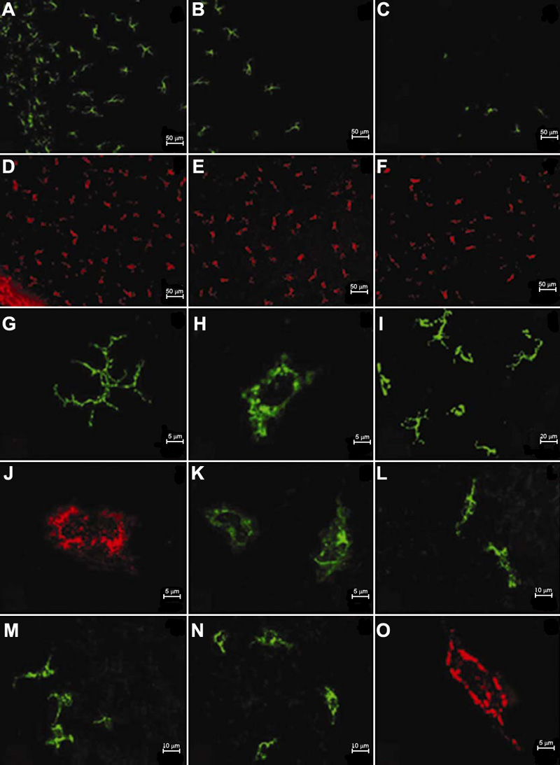
Figure 6. Distribution and morphology of labeled cells in the murine cornea at 24 h after intravitreal injection of fluorophore-conjugated reagents
A decreasing density of positive cells is observed from peripheral to central cornea (A,D: peripheral cornea, B,E: paracentral cornea, C,F: central cornea. A-C: MHC-II+ cells, D-F: OVA+ cells). Two populations of each antibody labeled cells, i.e., dendriform (G: MHC-II+ cell, I: CD11c+ cells) and round or irregular shapes (H: MHC-II+ cell, I: CD11c+ cells, J: F4/80+ cell, K: B7-1+ cell) are identified. F4/80+ cells seem to be more elongated in the peripheral area (L), slightly ramose in the paracentral region (M), and more round or oblong in the central cornea (N). All OVA+ cells show round, oblong or irregular shapes (O).