![]() Figure 4 of
Meng, Mol Vis 2007;
13:475-486.
Figure 4 of
Meng, Mol Vis 2007;
13:475-486.
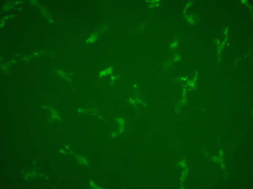
Figure 4. Intravital micrography of OVA+ cells in the murine cornea
OVA+ cells in the paracentral region of the murine cornea are detected by intravital microscopy 12 h after intravitreal injection (X200). The background is somewhat stronger.