![]() Figure 2 of
Meng, Mol Vis 2007;
13:475-486.
Figure 2 of
Meng, Mol Vis 2007;
13:475-486.
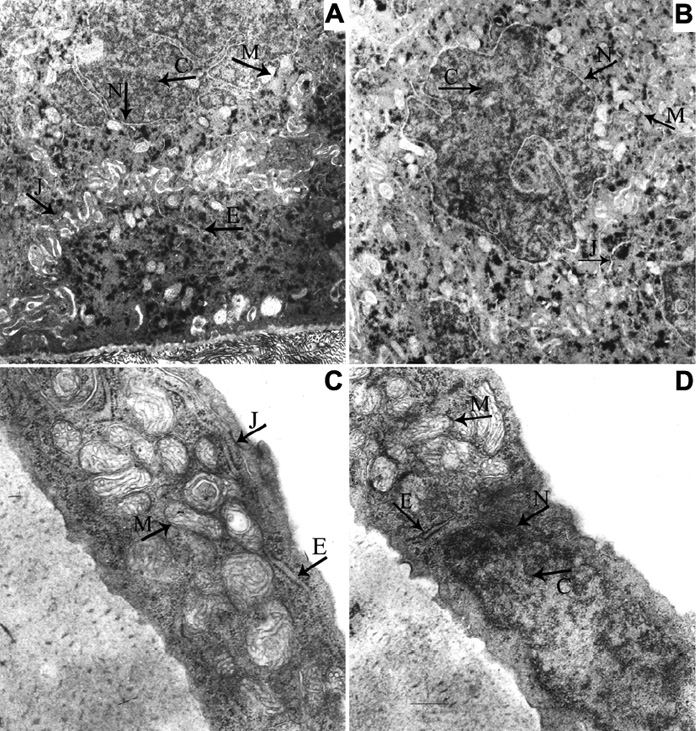
Figure 2. Ultrastructure of the cornea from the normal mouse and the mouse receiving intravitreal injection
Electron micrograph of the normal cornea (A: epithelium, C: endothelium) and the cornea of mouse receiving intravitreal injection (B: epithelium, D: endothelium). The mitochondrion has no ectasia, the mitochondrial outer and inner membranes are intact, and the mitochondria cristae arrange normally. The endoplasmic reticulum does not dilate. The nuclear membrane has integrity and the chromatin is distributed uniformly. The intercellular substance does not broaden and the cellular junctions were normal. The arrows indicate the location of different microorganelles. M: mitochondrion, E: endoplasmic reticulum, N: nuclear membrane; C: chromatin, J: cellular junction. The magnification in A and B is 12,000X while the magnification in C and D is 22,500X.