![]() Figure 10 of
Meng, Mol Vis 2007;
13:475-486.
Figure 10 of
Meng, Mol Vis 2007;
13:475-486.
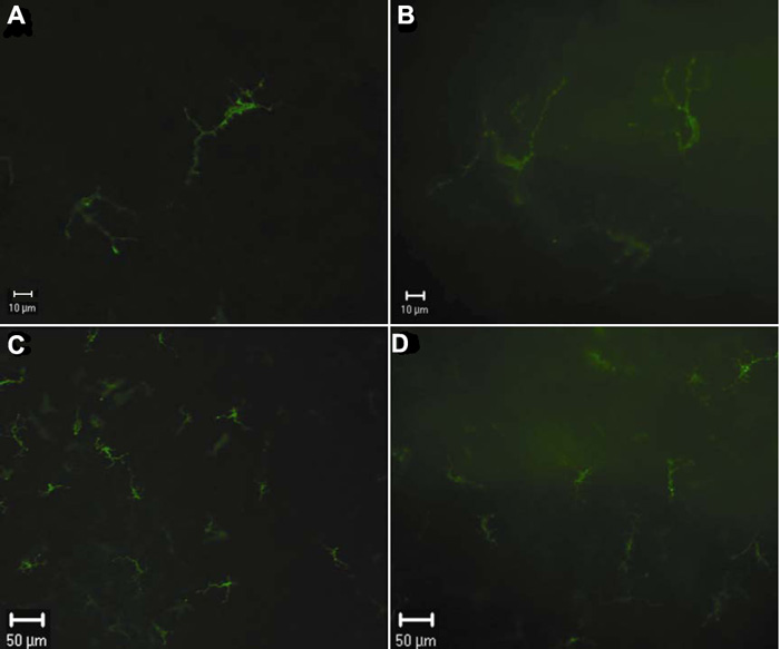
Figure 10. MHC-II positive cells stained in vivo and in vitro shown by epifluorescence microscopy
MHC-II+ cells stained in vivo 24 h after intravitreal injection (A and C) and in vitro (B and D) in the paracentral region of the murine cornea are observed by epifluorescence microscopy. The background in vitro staining is stronger than that in vivo staining. The labeled cells in vitro staining appear to be vaguely seen as compared with those in vivo staining.