![]() Figure 1 of
Meng, Mol Vis 2007;
13:475-486.
Figure 1 of
Meng, Mol Vis 2007;
13:475-486.
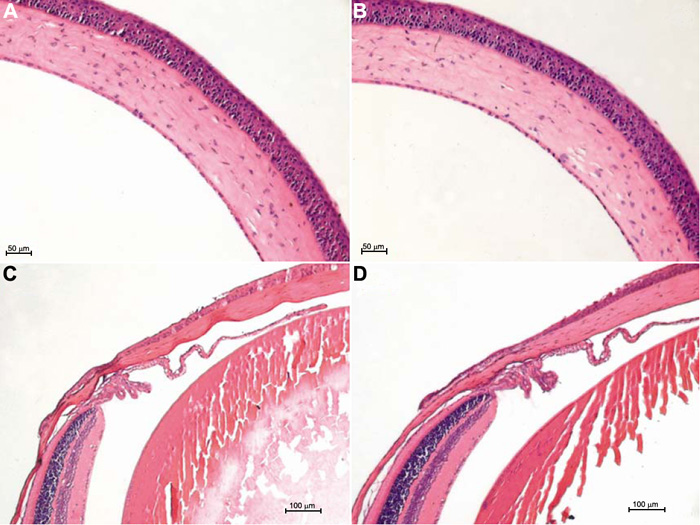
Figure 1. Histology of the cornea, iris-ciliary body, and retina in the normal mouse and the mouse receiving intravitreal injection
Light micrograph of the normal cornea, iris-ciliary body, and retina (A,C) and the cornea, iris-ciliary body, and retina of mice receiving intravitreal injection (B,D). The corneal epithelium, endothelium, and stroma are arranged in order and no inflammatory cells and edema are observed.