![]() Figure 1 of
Huang, Mol Vis 2007;
13:470-474.
Figure 1 of
Huang, Mol Vis 2007;
13:470-474.
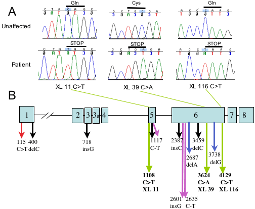
Figure 1. NHS mutation detection and localization
A: The identification of NHS nonsense mutations in three NHS families. Chromatograms from one affected and one unaffected individual are shown for each family. Above each tracing the base change is highlighted and the predicted stop codon is indicated. B: The localization of the fourteen reported NHS mutations within the NHS gene: red arrow [14]; black arrows [12]; green arrows (this study); purple arrows [15]; and blue arrows [13].