![]() Figure 7 of
Robertson, Mol Vis 2007;
13:457-469.
Figure 7 of
Robertson, Mol Vis 2007;
13:457-469.
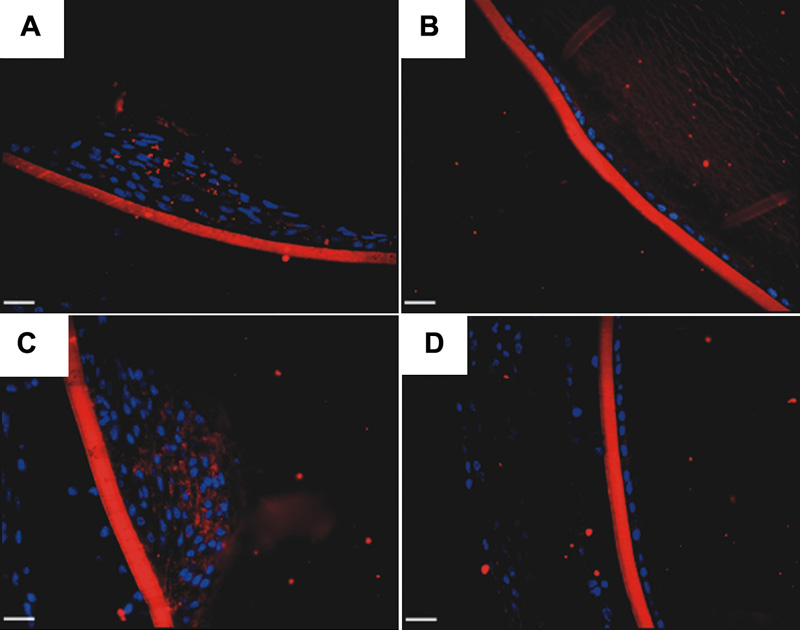
Figure 7. Collagen IV expression.
Immunolocalization of collagen IV was performed on paraffin sections of AdTGFβ1 (A and C) and AdDL (B and D) lenses on days 4 (A and B) and 21 (C and D). AdTGFβ1 treated eyes showed a marked accumulation of collagen IV in the plaques which was absent in epithelia of AdDL treated eyes. The red staining is collagen IV and the blue staining is DAPI. The scale bar is equal to 25 μm.