![]() Figure 6 of
Robertson, Mol Vis 2007;
13:457-469.
Figure 6 of
Robertson, Mol Vis 2007;
13:457-469.
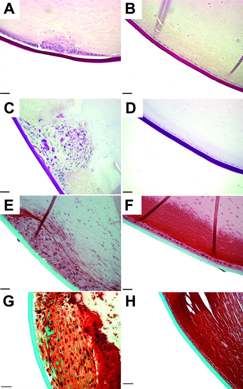
Figure 6. Matrix staining of AdTGFβ injected eyes.
Sections of AdTGFβ1 (A, C, E, and G) and AdDL (B, D, F, and H) lenses taken on days 4 (A, B, E, and F) and 21 (C, D, G, and H) were stained with PAS (A-D) to detect carbohydrates (purple) and Masson's trichrome (E-H) to detect collagens (green). AdTGFβ1 treated lenses showed accumulation of matrix which was barely detectable on day 4, but prominent on day 21. In contrast, AdDL treated lenses showed no matrix accumulation at any time point. The scale bar is equal to 25 μm.