![]() Figure 5 of
Robertson, Mol Vis 2007;
13:457-469.
Figure 5 of
Robertson, Mol Vis 2007;
13:457-469.
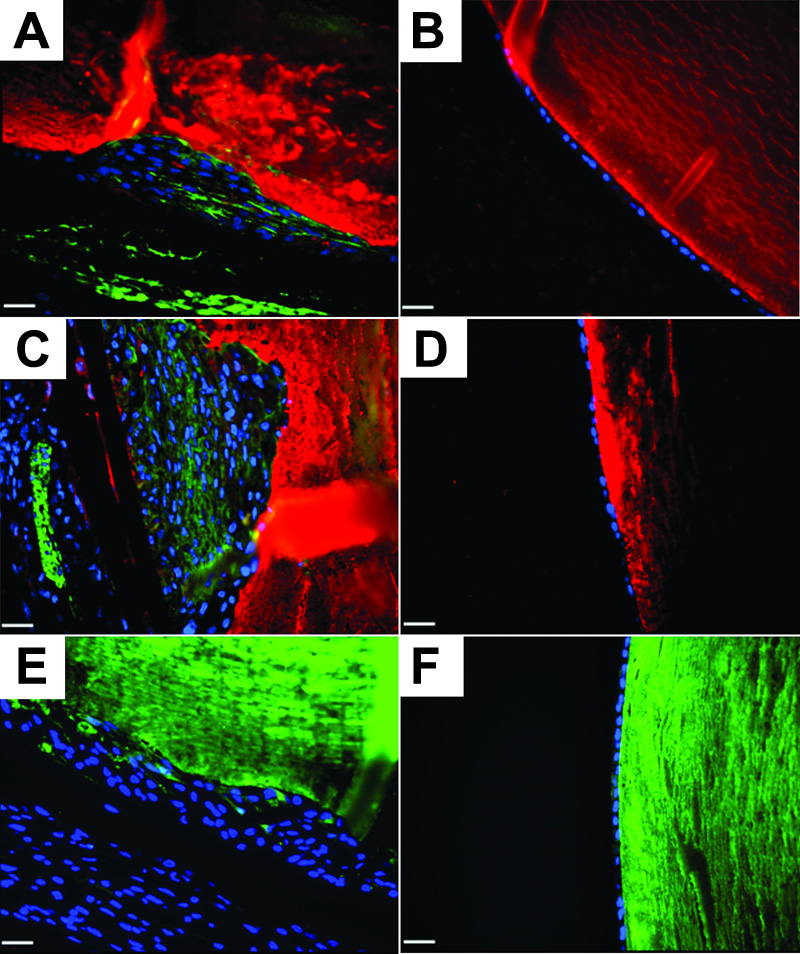
Figure 5. Crystallin expression in AdTGFβ1 treated eyes.
Sections were stained with anti-β (A-D; red) or anti-γ-crystallin (E and F; green) antibodies and counterstained with DAPI (blue). Additionally, β-crystallin stained sections were colocalized with αSMA (A-D; green). Sections were taken at four days (A and B) and at twenty one days (C-F) of both AdTGFβ1 (A, C, and E) and AdDL (B, D, and F). Both β- and γ-crystallin expression can be seen with in the plaques of AdTGFβ1 but not AdDL treated eyes. Epithelia of AdDL treated eyes showed no presence of crystallin or αSMA expression. The scale bar is equal to 25 μm.