![]() Figure 4 of
Robertson, Mol Vis 2007;
13:457-469.
Figure 4 of
Robertson, Mol Vis 2007;
13:457-469.
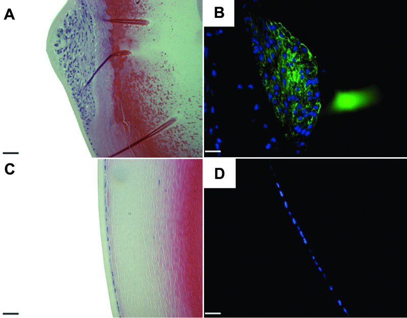
Figure 4. Effects of adenoviral gene transfer of active TGFβ1 after twenty one days.
Histological sections from C57 mice injected with AdTGFβ1 (A and B) or AdDL (C and D). Sections were stained with H&E (A and C) or subjected to immunostaining for αSMA (B and D). At 21 days post injection AdTGFβ1 treated eyes demonstrate large plaques which express a considerable amount of αSMA in contrast to AdDL treated eyes. The green stain is αSMA and the blue stain is DAPI. The scale bar is equal to 25 μm.