![]() Figure 3 of
Robertson, Mol Vis 2007;
13:457-469.
Figure 3 of
Robertson, Mol Vis 2007;
13:457-469.
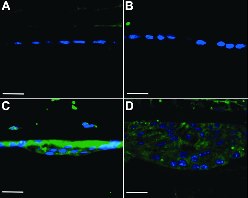
Figure 3. Immunolocalization of TGFβ1
Naïve (A), AdDL injected (B), and AdTGFβ1 injected eyes (C and D) were sectioned and subjected to TGFβ1 immunohistochemistry at 0 (A), 4 (B and C) and 21 (D) days after injection. Both naïve and AdDL eyes show no immunolocalization of TGFβ1 in the lens epithelium. Animals injected with AdTGFβ1 demonstrate prominent expression of TGFβ1 in the lens epithelium. The green stain is TGFβ1 and the blue stain is DAPI. The scale bar is equal to 25 μm.