![]() Figure 1 of
Robertson, Mol Vis 2007;
13:457-469.
Figure 1 of
Robertson, Mol Vis 2007;
13:457-469.
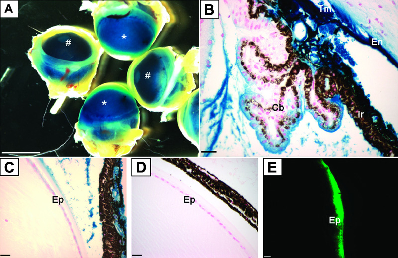
Figure 1. Expression of adenovirally transferred LacZ and GFP
Animals were injected with AdLacZ (A-C), AdDL (D), or AdGFP (E) intracamerally. Four days post injection eyes were removed and wholemount stained for LacZ activity (A) followed by paraffin sectioning (B,C,D) or frozen sectioning and visualized using a GFP filter (E). Blue corneal opacity (asterisks in A) indicates that cellular infection and production of transgene was successful. AdDL eyes remained clear (hash marks in A). B and C demonstrate transgene expression in the corneal endothelium (En), iris (Ir), ciliary body (Cb), trabecular meshwork (Tm), and lens epithelium (Ep). Transgene was absent in control vector treated eyes (D). Transgene expression is confirmed in the lens epithelium (Ep) using AdGFP (green). The scale bars in A = 1 mm, in B-D = 25 μm, and in E = 50 μm.