![]() Figure 5 of
Rosenthal, Mol Vis 2007;
13:443-456.
Figure 5 of
Rosenthal, Mol Vis 2007;
13:443-456.
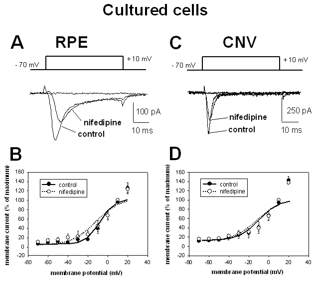
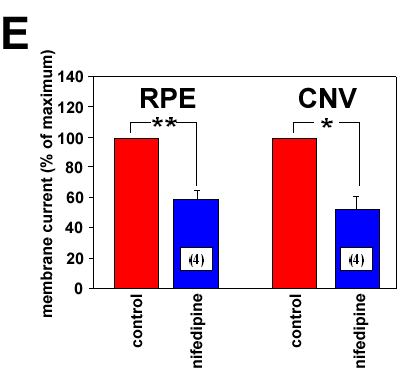
Figure 5. Effect of the dihydropyridine compound nifedipine on voltage-dependent Ba2+ currents of cultured retinal pigment epithelium cells
A: Ba2+ currents induced by a 50 ms voltage-step from -70 mV to +10 mV (upper panel) in a cultured retinal pigment epithelium (RPE) cell from an eye without choroidal neovascularization (CNV) under control conditions and in the presence of nifedipine (10-5 M). B: Curves of voltage-dependent activation from 14 cultured cells from eyes without CNV: 10 cells under control conditions and four cells in the presence of nifedipine. Currents were normalized to the maximal current amplitude and fitted using the Boltzmann equation. C: Ba2+ currents induced by a 50 ms voltage-step from -70 mV to +10 mV in a cultured RPE cell from a CNV membrane under control conditions and in the presence of nifedipine (10-5 M). D: Curves of voltage-dependent activation from 13 cultured cells from CNV membranes: nine cells under control conditions and four cells in the presence of nifedipine. Currents were normalized to the maximal current amplitude and fitted using the Boltzmann equation. E: Comparison of the inhibitory effect of nifedipine in cultured RPE cells from eyes without CNV and from CNV tissues. Maximal currents at +10 mV under control conditions were set as 100%.

