![]() Figure 4 of
Rosenthal, Mol Vis 2007;
13:443-456.
Figure 4 of
Rosenthal, Mol Vis 2007;
13:443-456.
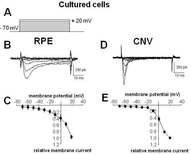
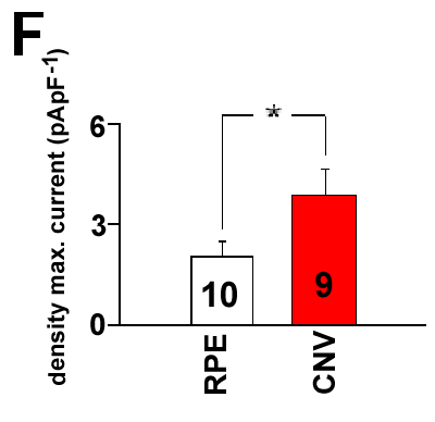
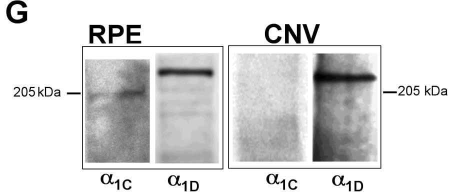
Figure 4. Voltage-dependent Ba2+ currents in cultured cells
A: Pattern of electrical stimulation to activate voltage-dependent Ba2+ currents. B: Ba2+ inward currents in a cultured human retinal pigment epithelium (RPE) cell of an eye without choroidal neovascularization (CNV). C: Current/voltage plot of currents from 10 cultured cells from eyes without CNV. D: Ba2+ inward currents in a cultured human RPE cell from CNV tissue. E: Current/voltage plot from nine cultured cells from CNV membranes. F: Comparison of maximal Ba2+ densities at +10 mV in cultured cells. G: Western blot of membrane proteins of cells from control eyes and CNV membranes. Left lane: staining for Cav1.2 (channel α1C) subunits; right lane: staining for Cav1.3 (channel α1D) subunits.