![]() Figure 2 of
Rosenthal, Mol Vis 2007;
13:443-456.
Figure 2 of
Rosenthal, Mol Vis 2007;
13:443-456.
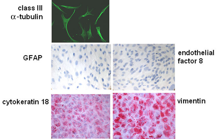
Figure 2. Analysis of retinal pigment epithelium cell cultures from CNV membranes
Sub-confluent cultures of retinal pigment epithelium (RPE) cells from choroidal neovascularization (CNV) tissues used for patch-clamp analysis were immunostained against class III β-tubulin. Confluent RPE cell cultures from CNV tissues were stained against glial acidic fibrillary protein (GFAP), endothelial factor 8, cytokeratin 18, or vimentin.