![]() Figure 5 of
Kokkinos, Mol Vis 2007;
13:418-430.
Figure 5 of
Kokkinos, Mol Vis 2007;
13:418-430.
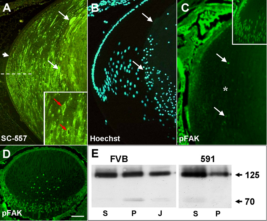
Figure 5. Expression of FAK in OVE591 transgenic lenses
A: Immunolocalization of FAK with the NH2-terminal antibody (SC557) in a P3 transgenic lens showing normal initiation of FAK expression in the posterior germinative zone (arrowhead) and expression in the transitional zone below the equator (dashed line). However, in more mature cortical and central fibers, FAK protein appears to aggregate abnormally in the cytoplasm and in perinuclear compartments (red arrows, inset) and is excluded from some nuclei. In the degenerate central lens fibers, intense reactivity for FAK is detectable in swollen fiber cells (arrows). B: Hoechst stained section shown in A showing where terminal differentiated fibers start to degenerate (arrows). C: Immunolocalization with the phosphoY397-FAK antibody on P2 lenses, just prior to appearance of the phenotype, showed greatly reduced expression of phosphorylated FAK in the fiber mass (asterisk) compared to wild-type lenses (inset). Some nuclear FAK is detected in nuclei of a few cortical fibers (arrows). D: In embryonic (E17.5) lenses, 7 days prior to phenotype initiation, expression of phospho-FAK is still present in the epithelium and fibers, similar to wild-type. E: Western blots of detergent-separated whole lens extracts (supernatant and pellet) from P3 FVB and OVE591 lenses, probed with SC558. In wild-type FVB lenses, equivalent amounts of 125 kDa FAK are present in the soluble (S) and in the cytoskeleton-associated (P) fractions, whereas in OVE591 lenses there was a shift of FAK from the cytoskeletal (P) to the soluble (S) fraction. A FAK-reactive band of approximately 70 kDa was evident in the P but not the S fraction of wild-type FVB lenses. In the 591 mutant lenses this band was absent from the P fraction but weakly detected in the S fraction. J indicates the Jurkat cell extract positive control. The scale bar in A-D indicate 100 μm; in A, inset, 200 μm; and in C, inset; 150 μm.