![]() Figure 2 of
Kokkinos, Mol Vis 2007;
13:418-430.
Figure 2 of
Kokkinos, Mol Vis 2007;
13:418-430.
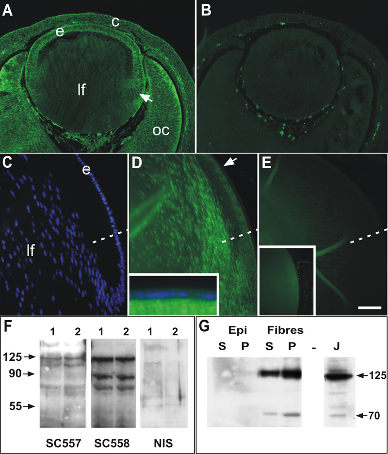
Figure 2. Immunolocalization of FAK in lens
Peptide antibodies to NH2-terminus (SC557) and COOH-terminus (SC558) domains of FAK were used to determine the presence of FAK protein in embryonic and postnatal lenses. A: Strong FAK reactivity in lens epithelium (e) and differentiating fibers (arrow) in E13.5 lens using the SC558 antibody. More mature fibers (lf) in the center of the lens showed little staining. Reactivity with this antibody was predominantly cytoplasmic and membrane-associated. Distinct staining was also detected in the optic cup (oc) and overlying cornea (c). B: SC558 pre-adsorbed with COOH-terminal peptide showed no reactivity in any ocular tissues; staining of blood cells is an autofluorescence artefact. C: Hoechst staining of section in D, showing epithelial (e) and fiber (lf) cells in the equatorial region of a P21 mouse lens. The equator is indicated by the dashed line in C-D. D: Distinct punctate reactivity for FAK (SC557 antibody) was detected in the epithelial cells of the posterior germinative zone (arrow) but not in anterior epithelial cells (inset). Reactivity was more intense in the transitional zone and in the cortical fibers and decreased in the more mature fiber cells. E: Non-immune serum controls showed no specific staining in sections from P21 lenses and preadsorption of SC557 with NH2-terminal peptide abolished almost all staining in the lens (inset). F: Western blots of whole lens extracts from two separate preparations probed with the SC557 and SC558 and non-immune rabbit IgG (NIS). SC557 revealed a distinct band at the predicted molecular mass of 125 kDa as well as lesser bands at about 110 kDa and about 65 kDa. SC558 also revealed the predicted 125 kDa as well as lower molecular weight species at about 85 kda and about 65 kDa. Membranes probed with the non-immune IgG showed no specific bands. G: Western blots of detergent-separated extracts (supernatant and pellet) from trimmed epithelial and fiber cell preparations (P9 rats), probed with the SC-558 antibody. FAK reactive bands at 125 and about 70 kDa were not detected in either soluble (S) or pellet (P) fractions from epithelial (Epi) cells, but were clearly detectable in both fiber cell preparations with more detectable in the cytoskeleton-associated fraction (P). J indicates Jurkat cell extract used as positive control. The scale bar in A and B represents 75 μm; in C-E, 50 μm; and in the insets, 125 μm.