![]() Figure 1 of
Kokkinos, Mol Vis 2007;
13:418-430.
Figure 1 of
Kokkinos, Mol Vis 2007;
13:418-430.
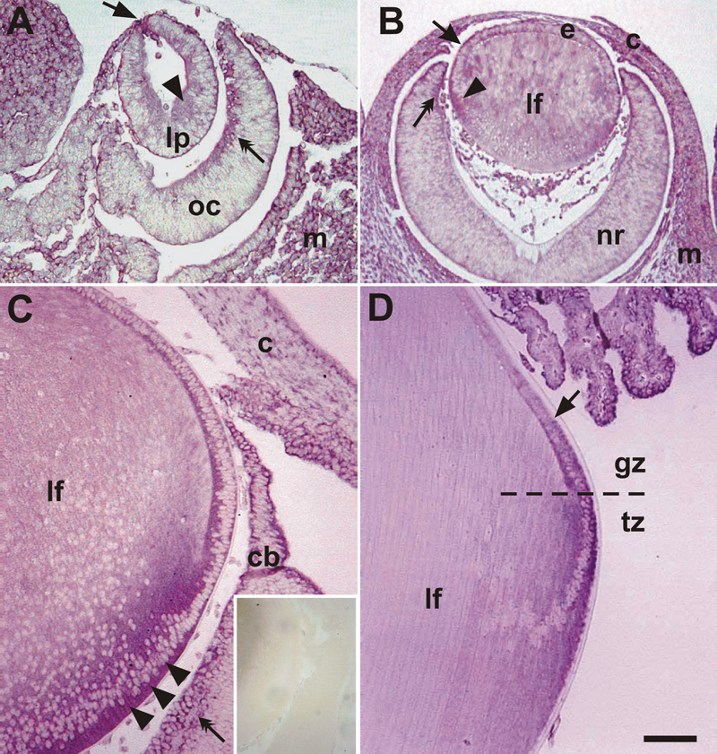
Figure 1. FAK mRNA expression during lens development
A: At E11.5, FAK was intensely expressed at the point of closure of the lens pit (arrow) with weaker expression detected at the apical cytoplasm of posterior cells of early pit/vesicle (arrowhead). Strong signals were also found along the inner optic cup (oc, double arrow) and in the extra-ocular mesenchyme (m). B: At E13.5, distinct FAK expression was detected in elongating fibers (arrowhead) of the late lens vesicle, while patchy expression (arrow) was detected in anterior epithelial cells (e). In the neural retina expression was predominantly found in the peripheral retina (double arrow) with little to no expression detected in the central regions. The developing cornea (c) and extra-ocular mesenchyme were also positive for FAK mRNA. C: At P1, FAK was expressed intensely in elongating fibers (lf), particularly in the basal cytoplasm (arrowheads). Weaker expression was detected in epithelial cells. Expression was also detected in the ciliary body (cb), corneal (c) epithelium, and ganglion cells of inner retina (double arrow). No staining was observed with the sense control probe (C, inset). D: At P21, strong FAK signal was detected in the lens transitional zone (tz) and posterior part (arrow) of the germinative zone (gz), whereas the more anterior epithelial cells and the mature fibers (lf) showed no expression. Strong expression was also detected in the ciliary body. Similar results were obtained with probes to 5' or 3' regions of the FAK cDNA. The scale bar in A represents 70 μm; in B, 150 μm; in C and D, 100 μm; and in C, inset, 300 μm.