![]() Figure 1 of
Ronkko, Mol Vis 2007;
13:408-417.
Figure 1 of
Ronkko, Mol Vis 2007;
13:408-417.
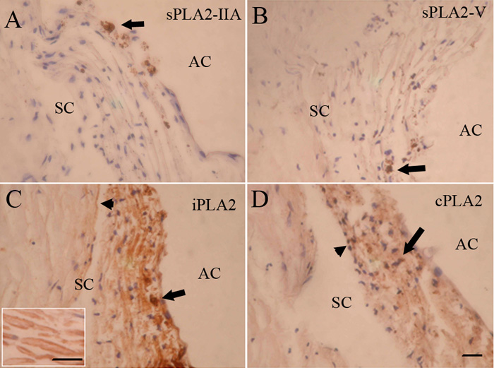
Figure 1. Immunohistochemical localization of PLA2s in normal human trabecular meshwork
Immunostaining for sPLA2-IIA (A) or sPLA2-V (B) was negative in the trabecular meshwork. Intense staining for sPLA2-IIA or sPLA2-V was evident in a few inflammatory-like cells (arrow). iPLA2 immunolabeling was strong (C). Labeling of the uveal and corneoscleral meshwork was stronger compared to the staining of the juxtacanalicular meshwork. Low positive staining was seen in the apical parts of the cells lining Schlemm's canal (arrowhead) as well as in nearby extracellular regions. Positive staining was also seen in a few macrophages (arrow). Inset: A portion of trabecular meshwork lamellae at higher magnification. Uveal trabecular meshwork cells covering the lamellae were more intensely labeled compared to connective tissue core. cPLA2 was weakly positive (D) and staining was slightly higher in uveal and corneoscleral meshwork compared to juxtacanalicular meshwork. The cells lining Schlemm's canal showed weak staining. Positive staining was seen in a few macrophages (arrow). AC, anterior chamber; SC, Schlemm's canal. The scale bar is equal to 50 μm.