![]() Figure 9 of
Xie, Mol Vis 2007;
13:397-407.
Figure 9 of
Xie, Mol Vis 2007;
13:397-407.
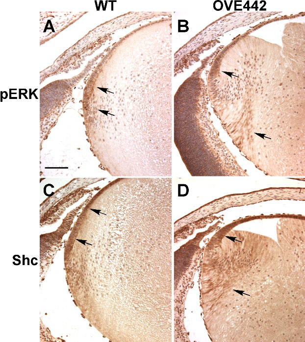
Figure 9. Immunohistochemical staining for pERK and Shc
In the WT E18.5 lens, pERK immunoreactivity was detected in the lens epithelial cells at the germinative zone and in the newly differentiating fiber cells (A, arrows). In the OVE442 transgenic lens, the pERK-staining area is expanded in the fiber cells (B, arrows). The immunostaining pattern for the adaptor protein Shc is also changed in the transgenic lens. In the WT lens, Shc is most strongly expressed in the lens epithelial cells (C, arrows). In the OVE442 transgenic lens, strong Shc staining was detected not only in the epithelial layer but also in the cortical fiber cells (D, arrows).