![]() Figure 6 of
Xie, Mol Vis 2007;
13:397-407.
Figure 6 of
Xie, Mol Vis 2007;
13:397-407.
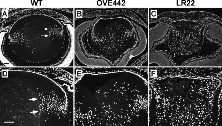
Figure 6. Fiber cell denucleation in newborn wild type (WT), OVE442 and LR22 mouse lenses
Newborn eye sections were stained with DAPI to reveal the distribution of cell nuclei. In the WT lens (A, D), fiber cell maturation is associated with denucleation and formation of a nuclear-free zone (arrows mark the margin of the area). In OVE442 (B, E) and LR22 (C, F) transgenic lenses, fiber cell nuclei are present across the entire lens section.