![]() Figure 5 of
Xie, Mol Vis 2007;
13:397-407.
Figure 5 of
Xie, Mol Vis 2007;
13:397-407.
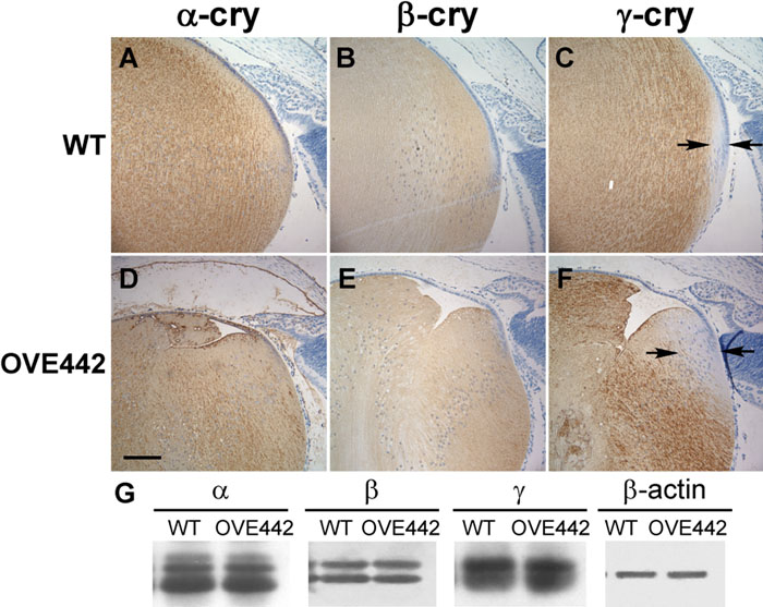
Figure 5. Crystallin expression detected by immunohistochemical staining and western blot
Eye sections of postnatal day 3 (P3) wild type (A-C) and OVE442 transgenic (D-F) mice were stained for α- (A, D), β- (B, E), and γ- (C, F) crystallins. In both genotypes, α-crystallin is expressed in all the lens cells, while β- and γ-crystallin are in the lens fiber cells. The space between the two arrows in (C) and (F) illustrate the area where γ-crystallin expression is absent. This area is expanded in the transgenic lens (F). With western blot analysis for α-, β-, and γ-crystallin (G), there was no detectable difference in the levels of crystallins between the wild type and transgenic mice. β-Actin was used as a control for protein loading.