![]() Figure 4 of
Xie, Mol Vis 2007;
13:397-407.
Figure 4 of
Xie, Mol Vis 2007;
13:397-407.
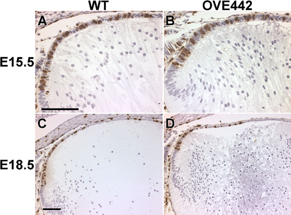
Figure 4. Cell proliferation in wild type (WT) and OVE442 transgenic mouse lenses
Lens cells at S-phase of the cell cycle were monitored with BrdU-labeling. BrdU-positive cells (brown nuclei) are distributed across the epithelial layer in the WT lenses (A, C) and OVE442 transgenic lenses (B, D). At E15.5 (A, B), the percentage of BrdU-positive nuclei is similar in the WT and transgenic lenses. At E18.5 (C, D), the percentage of BrdU-positive cells is slightly reduced in the transgenic lens, correlating with the phenotype of a reduced lens epithelial cell compartment.