![]() Figure 1 of
Xie, Mol Vis 2007;
13:397-407.
Figure 1 of
Xie, Mol Vis 2007;
13:397-407.
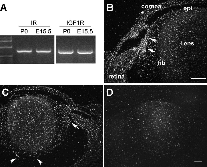
Figure 1. Expression of IR and IGF-1R in mouse lens
A: RT-PCR was used to detect IR and IGF-1R expression in embryonic day 15.5 (E15.5) and newborn (P0) mouse lenses. Expression patterns of IGF-1R (B) and IR (C) in newborn mouse lens were detected by in situ hybridization. IR sense probe was used as a negative control to monitor the background signal (D). Both IGF-1R and IR are expressed in the peripheral region of the lens (arrows in B and C). The IGF-1R signal was stronger than IR in the lens. Other tissues in the eye, including the cornea and retina, also express IGF-1R and IR. A high level of IR mRNA was found in the blood vessel cells surrounding the lens (arrowheads in C). Abbreviations used: epi, lens epithelial cells; fib, lens fiber cells. Scale bars in all the figures represent 100 μm.