![]() Figure 5 of
Chen, Mol Vis 2007;
13:374-387.
Figure 5 of
Chen, Mol Vis 2007;
13:374-387.
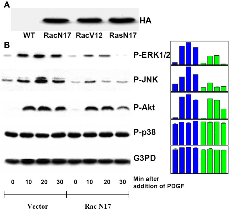
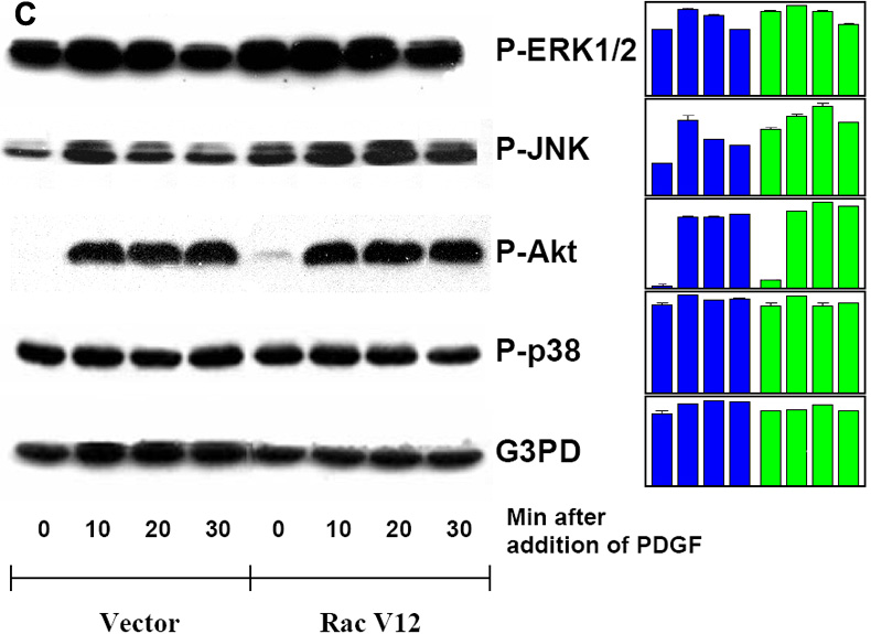
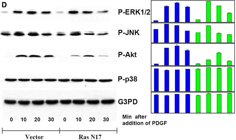
Figure 5. Western blot analysis of PDGF-activated MAP kinases in Ras or Rac transfected human lens epithelial B3 cells
Serum-deprived transiently transfected cells with and without inhibitor pretreatment were stimulated with PDGF (1 ng/ml) for 0, 10, 20, and 30 min, harvested and then lysed in lysis buffer. The vector-transfected cells were used as control. Cell lysates were applied on 10% SDS-PAGE, transblotted and probed with specific antibodies to HA, P-ERK1/2, P-JNK, P-Akt, P-p38, and G3PD. A: Wild type (WT) and transfected cells (Rac N17, Rac V12, Ras N17) were probed with anti-HA antibody. B: Vector- or Rac N17-transfected cells stimulated with PDGF were probed for P-ERK1/2, P-JNK, P-Akt, P-p38, and G3PD. C: Vector- or Rac V12-transfected cells stimulated with PDGF were probed for P-ERK1/2, P-JNK, P-Akt, P-p38 and G3PD. D: Vector- or Ras N17-transfected cells stimulated with PDGF were probed for P-ERK1/2, P-JNK, P-Akt, P-p38 and G3PD. The bar graph with averaged pixel values of the band intensities for each western blot is shown. Data presented are a typical representation of 2-3 separate experiments.